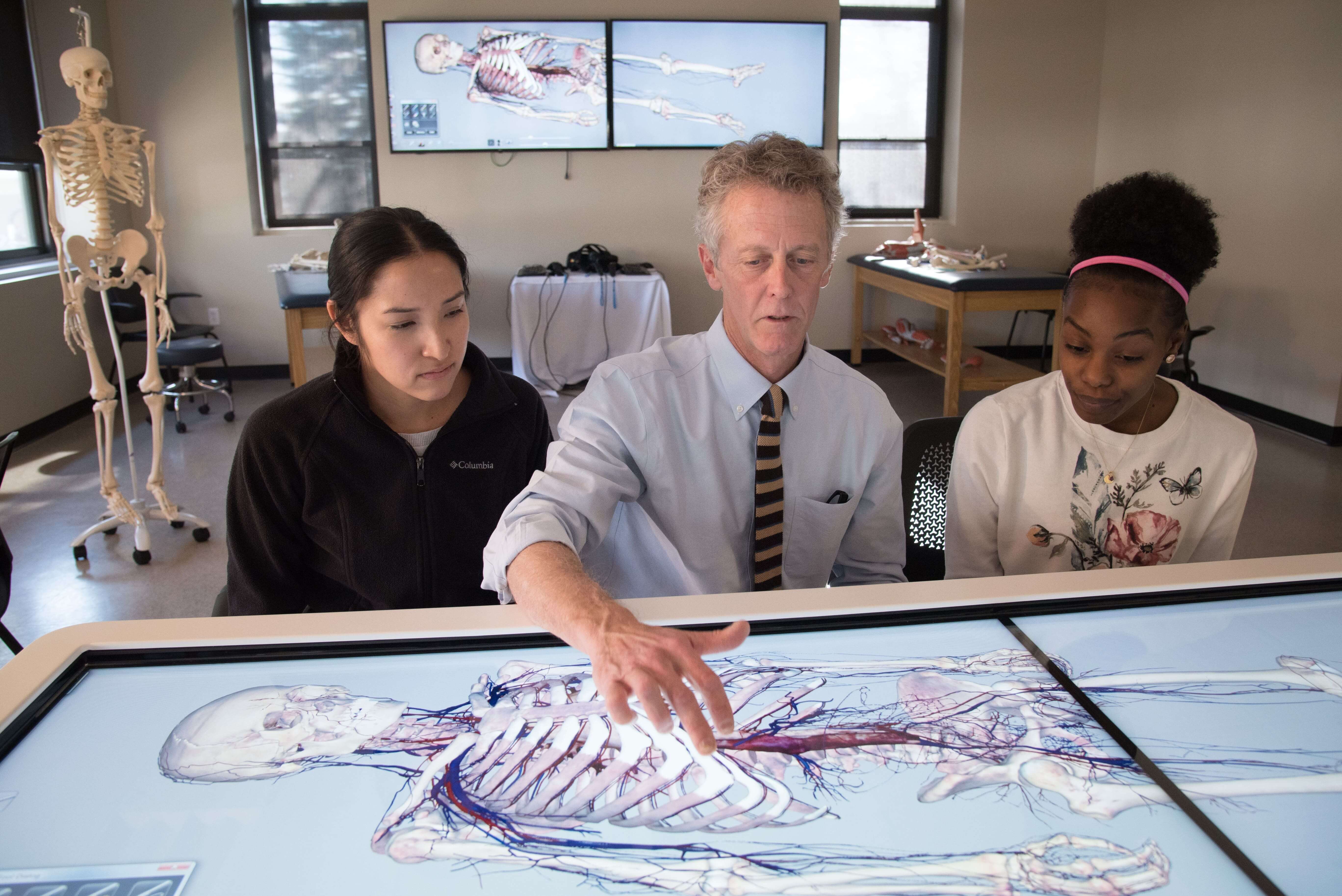
Unlocking the Wonders of Anatomy: Explore with a 3D Anatomy Table
A 3D anatomy table typically refers to a digital or physical educational tool that provides a three-dimensional representation of the human anatomy for learning and reference purposes. These tables are designed to help students, healthcare professionals, and researchers explore and understand the complexities of the human body in a more immersive and interactive way compared to traditional 2D anatomical images or textbooks.
Here are some
key features and components often found in 3D anatomy tables:
- 3D Visualization: The
primary feature is the ability to display anatomical structures in three
dimensions, allowing users to rotate, zoom in, and pan to view various
parts of the body from different angles.
- Interactive Interface:
Users can interact with the table through touchscreens, gestures, or
specialized input devices to explore anatomical structures, dissect
virtual cadavers, and access additional information about each structure.
- Layered Anatomy: Many
3D anatomy tables allow users to peel away layers of anatomy to reveal
deeper structures. This can be particularly useful for understanding the
relationships between different systems and organs.
- Labeling: Users can
often toggle labels on and off to identify and learn the names of specific
anatomical structures. This feature helps in memorizing anatomy.
- Functional Anatomy:
Some advanced tables can demonstrate the functions of muscles, organs, and
systems through animations and simulations, helping users understand how
the body works in real-time.
- Cross-Sectional Views: Users can view cross-sectional slices of the body to see internal structures in detail. This is especially valuable for medical imaging and surgery planning.
- Integration with Anatomy
Atlas: Many 3D anatomy tables are integrated with comprehensive
anatomy atlases and databases, providing access to detailed information,
clinical correlations, and pathology.
- Customization: Users
may be able to customize the display, focusing on specific regions or
systems of interest. This is beneficial for targeted learning or research.
- Compatibility: Some 3D
anatomy tables are designed for use with virtual reality (VR) or augmented
reality (AR) headsets, creating an even more immersive learning
experience.
- Education and Training:
3D anatomy tables are widely used in medical schools, healthcare
institutions, and research facilities for medical education, training, and
surgical planning.
These tables
can vary in terms of size and complexity, from small desktop versions to
larger, more advanced setups. Some popular digital dissection table systems include the Anatomage Table,
zSpace, and Touch of Life's 3D Human Anatomy Atlas. They continue to play a
significant role in modern medical education and healthcare by enhancing the
understanding of human anatomy and improving the training of medical
professionals.
A virtual dissection table, also known as a virtual anatomy
dissection table or virtual cadaver table, is an advanced educational and
medical technology that provides an immersive and interactive way to study and
dissect the human body virtually. These tables are primarily used in medical
schools, healthcare institutions, and research facilities to teach and explore
human anatomy without the need for physical cadavers.
Key features
and components of a virtual dissection table include:
- 3D Visualization: Similar to a 3D anatomy table, a virtual dissection table offers three-dimensional visualization of the human body. Users can manipulate and explore anatomical structures from various angles.
- Virtual Cadaver:
Instead of working with real cadavers, users interact with a digital
representation of a cadaver. This allows for repeated dissections without
the ethical concerns and logistical challenges associated with real
cadavers.
- Dissection Tools:
Users have access to virtual dissection tools such as scalpels, forceps,
and scissors. These tools enable them to virtually dissect and explore
different layers and structures within the body.
- Realistic Haptic Feedback:
Some advanced virtual dissection tables provide haptic feedback, allowing
users to feel resistance and tactile sensations when using dissection
tools. This enhances the realism of the experience.
- Layered Anatomy: Like
3D anatomy table India,
virtual dissection tables often allow users to peel away layers of anatomy
to reveal deeper structures. This feature helps in understanding the
relationships between different tissues and organs.
- Labeling and Information:
Users can access detailed information and labels about anatomical
structures, aiding in the learning process. Clinical and pathological
information may also be available.
- Cross-Sectional Views:
Virtual dissection tables can show cross-sectional views of the body,
helping users understand internal structures in detail, similar to medical
imaging techniques like CT scans and MRIs.
- Functional Anatomy:
Some virtual dissection tables include animations and simulations to
demonstrate the functions of muscles, organs, and systems.
- Customization: Users can often customize the dissection experience, focusing on specific regions or systems for targeted learning or research.
- Collaboration: In
educational settings, virtual dissection tables may support collaborative
learning, allowing multiple users to interact with the virtual cadaver
simultaneously.
Enquire Now
at: +91 8375931195
For More
information Please visit here: https://www.mavericsolution.com/


Comments
Post a Comment