Posts

Showing posts with the label virtual dissection table

How Hi-Fidelity Simulators and Virtual Dissection Tables Are Transforming Healthcare Education

Image
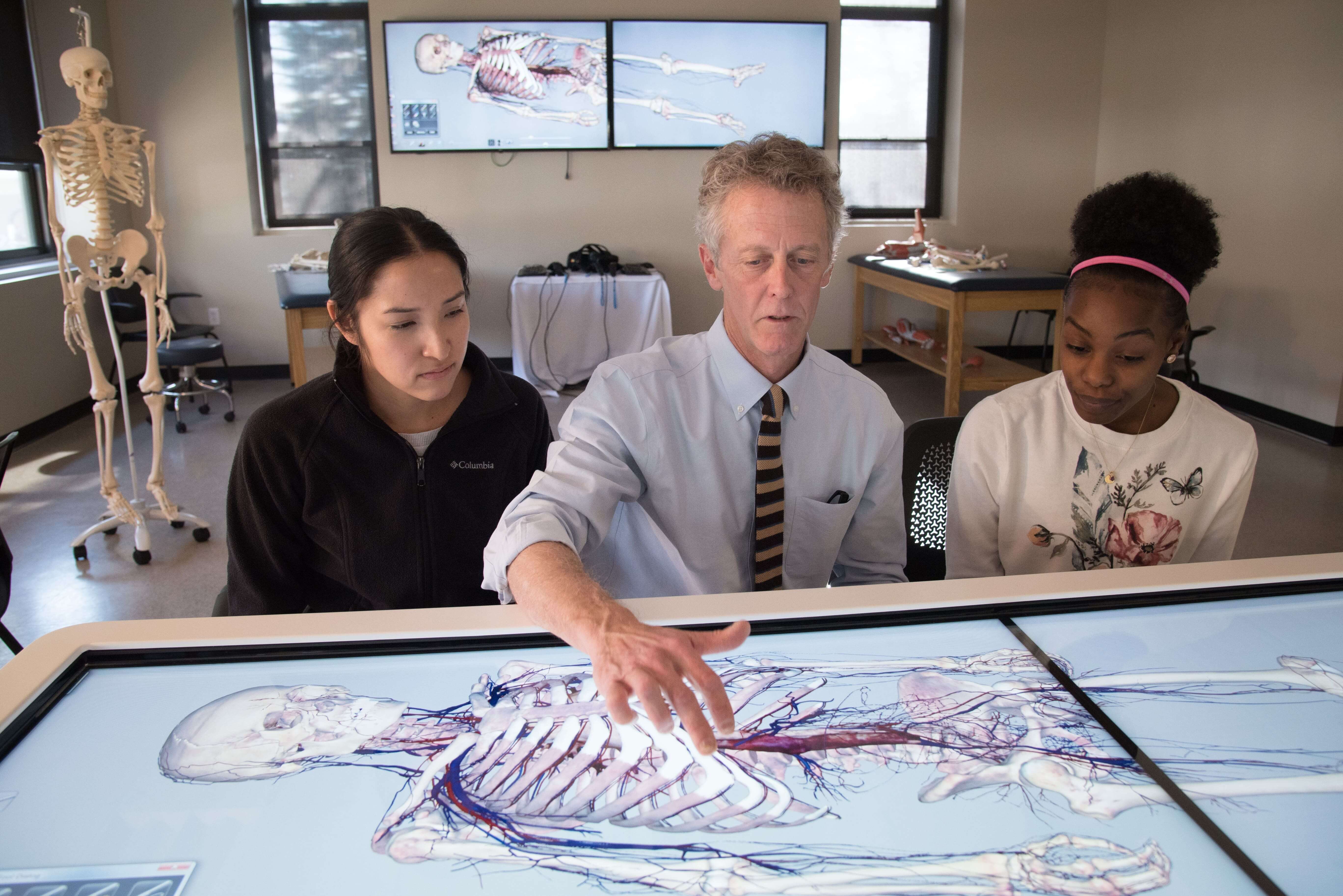
In an age where precision, safety, and real-time feedback are vital in medical education, advanced simulation technology has emerged as a game-changer. Tools like the hi-fidelity patient simulator , heart sound simulator , full OT simulator , and virtual dissection table are transforming the way future doctors, surgeons, and healthcare professionals learn and practice. This guide explores how each of these technologies contributes to improving healthcare training outcomes, reducing errors, and fostering confident clinical decision-making. 🔬 What is a Hi-Fidelity Patient Simulator? A hi-fidelity patient simulator is an advanced manikin-based simulation system designed to mimic real-life human physiological responses. Unlike low-fidelity models, these simulators respond to interventions such as CPR, incubation, drug administration, and even emotional cues like pain or anxiety. ✅ Core Features: Real-time physiological response (heart rate, BP, respiratory changes) Programmable...

Explore Human Anatomy Like Never Before with the Anatomage Table and Virtual Dissector

Image
 The field of medical education has undergone a profound transformation with the advent of advanced visualization technologies. Among the most groundbreaking innovations is the Anatomage Table  a cutting-edge virtual dissector that is redefining how anatomy is taught and understood in classrooms, hospitals, and research institutions around the world. What is Anatomage? Anatomage is a technology company known for developing one of the most advanced 3D anatomy visualization tools available today. Their flagship product, the Anatomage Table, is a life-sized, fully interactive anatomy table that allows users to virtually dissect a real human cadaver in incredible detail. With high-resolution images and precise anatomical accuracy, it provides a revolutionary learning experience for medical students, educators, and healthcare professionals. The Power of the Virtual Dissector Traditionally, anatomy was taught using physical cadavers, which often came with logistical, ethical,...

Medical Education in India with Virtual Dissection and 3D Anatomy Table

Image
The landscape of medical education in India is undergoing a transformative shift with the introduction of cutting-edge technologies like the   anatomy table ,   virtual dissection table , and   digital dissection table . As traditional cadaver-based learning faces ethical, logistical, and resource-related challenges, tools like the   3D anatomy table   and   Anatomage   have emerged as groundbreaking alternatives. What is an Anatomy Table? An  anatomy table  is a life-sized digital interface that simulates the human body in exquisite detail. These tables use advanced imaging technologies such as CT, MRI, and real anatomical data to allow students to interactively explore human anatomy. Unlike traditional cadaveric dissections, anatomy tables provide a clean, ethical, and repeatable learning experience. In India, where medical colleges often deal with cadaver shortages and increasing student populations, the adoption of virtual dissection tool...

Medical Education The Rise of Virtual and 3D Anatomy Tables in India

Image
Anatomy has always been the cornerstone of medical education, helping students understand the structure and function of the human body. Traditionally, this involved dissecting cadavers, a practice that, while essential, has its ethical, practical, and logistical limitations. With advancements in technology, the emergence of digital and virtual anatomy tables has revolutionized the way medical students and professionals engage with human anatomy. These innovations provide a more interactive, immersive, and educationally rich experience for those studying the human body, particularly in countries like India, where such advancements are gaining significant traction. Anatomy Table: A Game-Changer for Medical Education The anatomy table , in its most advanced form, has become an indispensable tool in modern medical institutions. A digital or virtual dissection table allows students to explore the human body in detail without the need for traditional cadaveric dissection. These tables integr...

Unveiling the Virtual Anatomy Dissection Table- A Journey through the Digital Realm of Anatomy

Image
In the ever-evolving landscape of medical education, innovation stands as a beacon, guiding both students and educators toward enhanced learning experiences. Among the myriad advancements, the    Anatomy Table   emerges as a revolutionary tool, transcending traditional methods and propelling anatomical exploration into the digital realm. Picture this a sleek, state-of-the-art table adorned with a high-definition touchscreen interface, beckoning curious minds to embark on a journey through the intricate landscape of the human body. No scalpels or formaldehyde-tinged specimens here. Instead, students are invited to delve into a meticulously crafted virtual environment where anatomy comes to life with unparalleled vividness.   Gone are the days of static textbooks and two-dimensional illustrations. The Virtual Anatomy Dissection Table offers an immersive, interactive experience that transcends the confines of traditional learning. With a simple swipe of a finger, users ...

Unlocking the Wonders of Anatomy: Explore with a 3D Anatomy Table

Image
A 3D anatomy table typically refers to a digital or physical educational tool that provides a three-dimensional representation of the human anatomy for learning and reference purposes. These tables are designed to help students, healthcare professionals, and researchers explore and understand the complexities of the human body in a more immersive and interactive way compared to traditional 2D anatomical images or textbooks. Here are some key features and components often found in 3D anatomy tables: 3D Visualization: The primary feature is the ability to display anatomical structures in three dimensions, allowing users to rotate, zoom in, and pan to view various parts of the body from different angles. Interactive Interface: Users can interact with the table through touchscreens, gestures, or specialized input devices to explore anatomical structures, dissect virtual cadavers, and access additional information about each structure. Layered Anat...