Posts

Showing posts with the label real tissue dissection table

Why Every Medical Institution Needs a 3D Anatomy Table?

Image
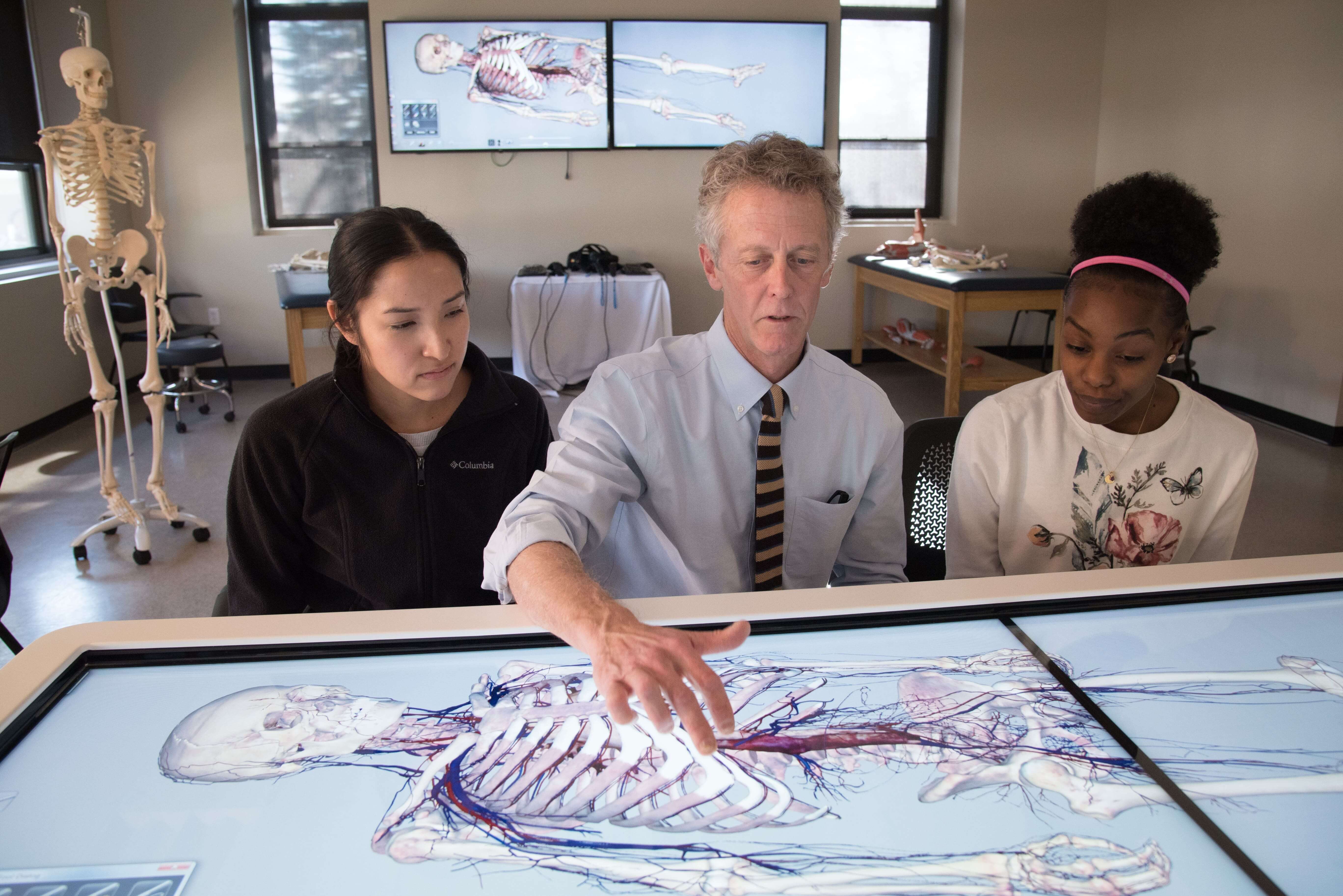
The study of human anatomy has always been the backbone of medical education. For centuries, students relied on cadavers, textbooks, and static models to learn the complex structure of the human body. While these methods have laid the foundation for medical learning, they often fall short in providing the depth and flexibility that modern healthcare demands. This is where the 3D anatomy table has changed the landscape of medical education and training. A 3D anatomy table is a high-tech interactive system that allows users to explore the human body in a way that traditional methods simply cannot. Instead of a flat image or limited model, this tool provides a lifelike, full-scale digital representation of human anatomy. Students, doctors, and researchers can rotate, dissect, and zoom into different layers of the body, gaining a level of understanding that is far more detailed than traditional learning aids. A Modern Solution for Medical Training One of the biggest advantages of the a...

Why the Anatomage Table is Changing How India Learns Human Anatomy?

Image
The Anatomage Table is a revolutionary tool that provides a virtual dissection experience with incredibly life-like visuals. With its high-resolution, 3D representation of real human anatomy, it allows users to explore the human body layer by layer, organ by organ, and system by system—without ever needing a scalpel. This makes it not only a powerful learning tool but also a safer and more sustainable alternative to traditional dissection methods. In India , medical colleges, universities, and healthcare training centers are increasingly adopting the Anatomy Table India edition to enhance their teaching methodologies. The table supports interactive and engaging learning, making complex anatomical concepts easier to grasp. Students can rotate the virtual cadaver, zoom in on specific organs, remove layers of tissue, and even simulate clinical conditions. These features give learners a more detailed and accurate understanding of the human body compared to traditional methods. While cad...

Virtual Anatomy Dissection Tables vs. Real Tissue Dissection Medical Education with Anatomage and Virtual Dissectors

Image
In recent years, advancements in medical education technology have led to a shift from traditional dissection methods to virtual anatomy dissection tables, such as those provided by Anatomage. This transition is reshaping how students, educators, and professionals engage with human anatomy. While real tissue dissection tables remain an essential part of medical training, virtual dissectors offer unique advantages that enhance learning and accessibility. The Rise of the Virtual Anatomy Dissection Table A  virtual anatomy dissection table  is a digital tool that provides an interactive platform for studying human anatomy without the need for physical cadavers. These tables allow users to explore the body layer-by-layer, viewing muscles, bones, organs, and blood vessels in precise detail. Anatomage, a pioneer in the field, has developed sophisticated tables that replicate human anatomy with high accuracy and offer an immersive experience for medical students and professionals. Th...

Unveiling the Virtual Anatomy Dissection Table- A Journey through the Digital Realm of Anatomy

Image
In the ever-evolving landscape of medical education, innovation stands as a beacon, guiding both students and educators toward enhanced learning experiences. Among the myriad advancements, the    Anatomy Table   emerges as a revolutionary tool, transcending traditional methods and propelling anatomical exploration into the digital realm. Picture this a sleek, state-of-the-art table adorned with a high-definition touchscreen interface, beckoning curious minds to embark on a journey through the intricate landscape of the human body. No scalpels or formaldehyde-tinged specimens here. Instead, students are invited to delve into a meticulously crafted virtual environment where anatomy comes to life with unparalleled vividness.   Gone are the days of static textbooks and two-dimensional illustrations. The Virtual Anatomy Dissection Table offers an immersive, interactive experience that transcends the confines of traditional learning. With a simple swipe of a finger, users ...

Unlocking the Wonders of Anatomy: Explore with a 3D Anatomy Table

Image
A 3D anatomy table typically refers to a digital or physical educational tool that provides a three-dimensional representation of the human anatomy for learning and reference purposes. These tables are designed to help students, healthcare professionals, and researchers explore and understand the complexities of the human body in a more immersive and interactive way compared to traditional 2D anatomical images or textbooks. Here are some key features and components often found in 3D anatomy tables: 3D Visualization: The primary feature is the ability to display anatomical structures in three dimensions, allowing users to rotate, zoom in, and pan to view various parts of the body from different angles. Interactive Interface: Users can interact with the table through touchscreens, gestures, or specialized input devices to explore anatomical structures, dissect virtual cadavers, and access additional information about each structure. Layered Anat...